
The wave of digitalization has been sweeping through the field of dentistry for over a decade, and intraoral scanners have gradually evolved from novelties into standard equipment in many clinics. Yet, a look back at their adoption reveals an intriguing phenomenon: what truly fosters a dentist’s long-term trust in a scanner is often not the headline-grabbing accuracy figure or the frame-per-second scanning speed, but rather the nuanced details of the user experience—the balance of the device in hand, the temperature of the scanner tip upon entering the mouth, and the reliability of the lens remaining clear throughout lengthy procedures. It is precisely these seemingly subtle design considerations that determine whether a scanner can truly integrate into daily clinical practice and become a natural extension of the dentist’s hand.
I. The “Silent Variables” in the Clinic: Efficiency Black Holes Long Overlooked
In the hustle of clinical practice, breakdowns in digital workflows often occur at the most inconspicuous points. Scanner tips fog up due to the temperature difference between the oral cavity and the outside air, forcing the clinician to pause, remove and wipe the tip, and then re-insert it; cables get stepped on or tugged by chairside equipment, causing loose connections that interrupt data transmission and trigger software reconnection prompts; sterilization protocols for scanner tips are cumbersome, requiring specialized tools for disassembly, leaving nurses scrambling during the brief intervals between patient appointments. These seemingly trivial annoyances never appear on product comparison charts, yet they steadily drain the clinical team’s time and energy, accumulating into dozens of workflow interruptions and efficiency losses every day.
These are precisely the “silent variables” in equipment design that have long been overlooked. No matter how fast a scanner’s theoretical speed or how high its precision, if the clinician must stop every three teeth to deal with fogging or check cable connections, overall clinical efficiency suffers significantly, and the clinician’s workflow—their “flow state”—is constantly disrupted. Conversely, when every detail of the device anticipates potential clinical disturbances and offers preemptive solutions, the scanning process achieves a seamless, almost imperceptible fluidity. This fluidity is, in itself, the best guarantee of scanning accuracy and data integrity, as every restart following an interruption carries the risk of positional misalignment and stitching errors.
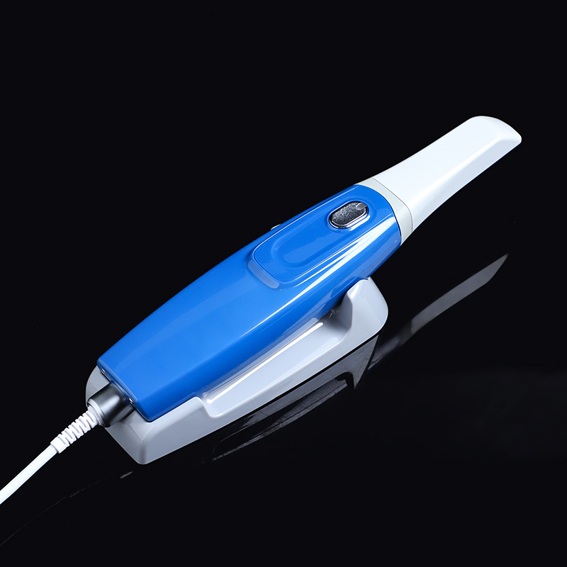
The design philosophy of the XANGTECH XT-OS33 is rooted in a deep understanding of these clinical “silent variables.” Its built-in intelligent heating system rapidly warms up upon startup and maintains a constant temperature. On one hand, it preheats the tip surface to prevent instant fogging caused by temperature differentials upon entering the mouth, allowing clinicians to complete a full-arch scan without repeatedly wiping the lens. On the other hand, the heated tip closely matches human body temperature upon contact with oral soft tissues; this significantly enhances comfort—especially for children and sensitive patients—by effectively reducing aversion and gag reflexes triggered by cold sensations, thereby making the scanning process much smoother.
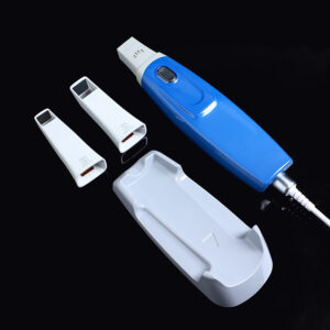
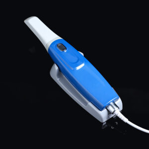
The design of the scanner tip also reflects a profound consideration for clinical efficiency. The XT-OS33 features a detachable scanner head and handle, allowing for replacement within three seconds without the need for tools. After a scan, the nurse can simply remove the used head for soaking disinfection or autoclave sterilization while attaching a new, sterile head to the handle, readying the device for the next patient. This plug-and-play, rapid-turnaround design significantly reduces room preparation time and meets the diverse infection control standards of various clinics. Moreover, the compact scanner head—combined with a mirror angle of nearly 45°—enables easy access to the posterior tooth region, narrow spaces, and the mouths of pediatric patients; it successfully captures complete data from areas that are typically “blind spots” in scanning, such as the lingual surfaces of anterior teeth and the distal surfaces of molars, thereby greatly expanding the device’s clinical utility.
II. From Data Acquisition to Collaborative Decision-Making: A Communication Revolution Driven by High-Fidelity Color Imaging
Initially, the core mission of intraoral scanners was singular: to capture digital impressions with sufficient precision to replace the cumbersome process of traditional silicone impression-taking. However, the appearance of high-definition color 3D models on clinic screens ushered in an unexpected shift—transforming the technology into a new language for clinical communication that fundamentally reshaped the dialogue between dentist and patient.
In dental practice, patients often struggle to interpret the radiolucent areas and 2D grayscale images found on X-rays, let alone visualize the conditions deep within their own mouths. When dentists use technical terms like “proximal caries,” “wedge-shaped defects,” or “cervical abrasion,” patients often lack the corresponding visual concepts, forcing them to rely on imagination—and the gap between that imagination and reality is frequently the root cause of misunderstandings. In contrast, color 3D models offer realistic color reproduction and three-dimensional form, clearly displaying details such as surface discoloration, inflamed gingival margins, the marginal integrity of existing restorations, and the distribution of dental plaque. Dentists no longer need to struggle with metaphors and gestures to explain the rationale for a procedure or the exact nature of a problem; patients need only look at the screen to see the conditions in their mouths firsthand.
This shift—moving from “relying on professional judgment” to “witnessing the facts”—significantly reduces uncertainty and psychological resistance during the decision-making process. When patients clearly see dark fissures on the occlusal surfaces of posterior teeth, pigmented plaque buildup on the lingual surfaces of anterior teeth, or distinct groove-like defects at the gumline, the necessity of treatment becomes self-evident. Patient understanding and acceptance of treatment plans improve markedly, and the trust between dentist and patient is substantially strengthened through this transparent communication.
The XT-OS33’s performance in high-fidelity color imaging provides the solid technical foundation for this communication revolution. Precisely calibrated at the factory, the device requires no periodic maintenance or additional adjustments during clinical use; it is ready to generate 3D models with accurate color reproduction and rich textural detail straight out of the box. Dentists can use the 3D color images displayed on the screen as a communication tool to point out the specific condition of each tooth—such as the depth and extent of cavities, the level of plaque accumulation, and the distribution of gingival inflammation—thereby transforming abstract medical diagnoses into visual information that patients can intuitively grasp and significantly reducing the cognitive gaps often caused by verbal descriptions. This positioning elevates the XT-OS33 beyond a mere data acquisition device, transforming it into a central communication hub within the clinic that fosters consensus between dentist and patient and increases treatment acceptance.
III. Laying the Foundation for an Ecological Connection: A Stable and Reliable Long-Term Partner
The ultimate form of digital dentistry is inevitably a seamless, closed-loop system spanning scanning, design, manufacturing, and clinical delivery. However, the reality for most clinics is that data silos between different equipment brands remain, and the adoption of cloud-based collaboration varies; many clinics are still in the transitional phase of moving from traditional workflows to digital ones. Against this backdrop, the true value of a scanner lies not merely in the tasks it can currently perform, but in its ability—through sufficient stability, reliability, and openness—to serve as a solid starting point for a clinic’s journey toward a fully digital workflow.
This means that factors such as compatibility with mainstream design software, support for future functional upgrades via interface protocols, and the hardware’s inherent reliability for long-term use—issues that might seem abstract—ultimately determine the scanner’s lifespan and return on investment. Devices that are permanently calibrated at the factory and require no periodic clinical maintenance fundamentally reduce operational burdens and hidden costs for clinics. This allows practitioners to focus entirely on patient care rather than on daily equipment upkeep or technical support coordination.
The XT-OS33 embodies this pragmatic yet forward-looking design philosophy. It utilizes a direct connection method—eliminating the need for external adapters or complex power configurations—which simplifies the chairside setup and keeps the workspace tidy and organized. The user interface currently supports 15 languages, including Polish, Spanish, and Russian, covering major markets across Europe, the Americas, and Asia-Pacific. This significantly lowers training costs and barriers to entry for staff in multilingual clinics or multinational medical institutions, enabling rapid integration into clinical environments across diverse regions. Furthermore, the XT-OS33’s open software architecture and standardized data output formats provide ample scope for future applications, such as cloud-based case management, AI-assisted diagnosis, and digital restoration design. When a clinic is ready to transition to a fully digital workflow, the XT-OS33 serves as a reliable data entry point, seamlessly connecting to subsequent design and manufacturing stages and avoiding the sunk costs associated with replacing an entire system due to equipment incompatibility.
IV. Seamless Human-Machine Integration: Technology That Fades into the Background
Intraoral scanning is a delicate procedure requiring the scanner tip to constantly adjust its angle within the confined space of the mouth to capture every axial surface of each tooth. In complex cases, a single scan can last several minutes or longer, demanding that the clinician maintain a steady hand and a fluid scanning path throughout. These operational requirements place high demands on the device’s weight distribution and ergonomics: excessive weight accelerates hand fatigue, leading to path deviations and the accumulation of stitching errors, while a poorly balanced center of gravity creates unwanted torque during rotation, making the procedure more strenuous.
The XT-OS33 features a systematically optimized ergonomic design. Weighing just 210 grams, the device boasts a streamlined profile that conforms to the natural curve of the palm, ensuring even pressure distribution and allowing for stable control without excessive finger strain. Even when handling multiple consecutive cases, the clinician’s hand muscles remain relaxed, preventing the soreness or stiffness often associated with prolonged use. This comfortable, natural grip translates directly into greater stability in the scanning path and continuity in data acquisition; clinicians can adjust angles with ease and precisely control the scanner tip’s movement, resulting in more complete and accurate digital models. When the tool effectively “fades into the background” through its seamless feel, the clinician can focus their full attention on the patient’s teeth and oral tissues—the ideal state of human-machine integration.
Technological advancements in intraoral scanners have never ceased; iterations bringing higher frame rates, reduced error margins, and greater depth of field continue to emerge. Yet, what truly propels digital dentistry from being merely “functional” to “user-friendly”—and from a “laboratory concept” to “everyday clinical practice”—is not merely innovation at the level of algorithms and sensors. Rather, it is the device’s profound understanding of, and precise response to, every genuine need within the clinical environment.
When a scanner eliminates fogging issues with temperature-controlled lenses, prevents cable disconnection via threaded locking interfaces, reduces clinician fatigue with a lightweight, balanced handpiece, simplifies sterilization workflows with detachable scanning tips, bridges the gap between doctor and patient through high-fidelity color models, and accommodates diverse markets and future applications with a multilingual interface and open architecture—it ceases to be a cold, impersonal tool requiring the clinician to “adapt” or “compromise.” Instead, it becomes a reliable partner that truly understands the clinical workflow, the clinician, and the patient.
The XT-OS33 embodies precisely this evolutionary path: eschewing flashy hype and gimmicky specifications detached from clinical reality, it instead earns the long-term trust of clinical professionals by steadfastly resolving every “minor inconvenience” with precision. While accuracy serves as the cornerstone of digital dentistry, it is “temperature”—a design ethos that ensures patient comfort, clinician ease of use, and seamless communication—that breathes life into the technology. The future of digital dentistry belongs to those creators who can both master micron-level precision and heed every genuine need expressed within the dental clinic.


